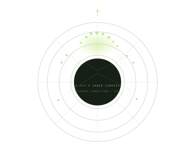
Find the bright patch.
Each glow is a neuron’s simulated activity. Together, a patch of active compass neurons represents a direction.
 INNER COMPASS
SMALL BRAINS. EXTRAORDINARY STRUCTURE.
Somewhere inside a fly’s brain, a small circuit remembers which way it’s facing. Now you can step inside it.
REAL FLYWIRE DATA · INTERACTIVE 3D · NO INSTALL NEEDED
A small window into
the architecture of navigation.
THE LIVE EXPERIMENT
Orbit the circuit. Turn the fly.
Discover what happens when the wiring changes.
Drag to orbit · Pinch to zoom · Tap a neuron
A FIELD GUIDE TO YOUR FLY
You don’t need to be a neuroscientist.
Just bring a little curiosity.
Each glow is a neuron’s simulated activity. Together, a patch of active compass neurons represents a direction.
Orbit a schematic ring, or explore reconstructed neuron branches. Both views follow the exact same live simulation.
Turn, then center the slider. Switch the connections to random destinations and watch the patch lose its shape.
↳ A little structure makes a world of difference.
This is a simplified rate model on a measured FlyWire subgraph, not a validated reconstruction of a living fly. Every neuron updates from its connected inputs. The same dynamics and input are used in both modes.
Connection weights start as synapse counts, then receive population-specific normalization and gains. Acetylcholine is modeled as excitatory; GABA and glutamate as inhibitory. Glutamate’s effect depends on receptors, so its sign here is an assumption. Four cells with unresolved transmitter predictions are excluded.
Turning supplies a moving, modeled heading cue; this demo does not claim to reproduce biological angular-velocity integration. This cue can organize either mode, so center the slider to compare persistence. At rest the cue switches off, and real wiring can drift toward preferred headings. Ring positions are inferred from connectivity, not measured anatomical coordinates. Zero degrees is arbitrary.
Shuffling permutes each neuron’s outgoing destinations, including empty slots. This preserves the exact edge count, outgoing weights, and transmitter sign, while breaking spatial organization. Patch coherence is the circular concentration of EPG activity; it is not biological accuracy.
The 3D ring is a schematic. The anatomy view uses simplified skeletons of these neurons from the published FlyWire v783 reconstruction. Skeleton branches show cell shape, not individual synaptic connections. Activity is assigned per neuron, not simulated separately along each branch. Anatomy stays fixed when wiring is shuffled.
FlyWire connectome ↗ · The biological ring attractor ↗ · Anatomy source ↗ · Circuit provenance ↗ · Anatomy provenance ↗